
CBCT Scan
Cone Beam Computed Tomography (CBCT) scan
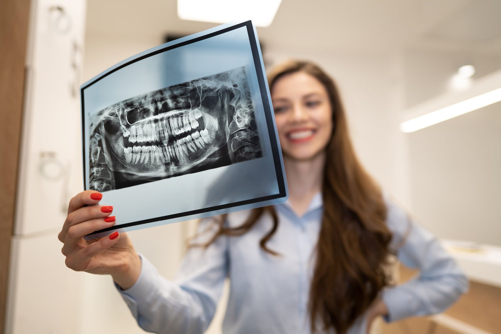
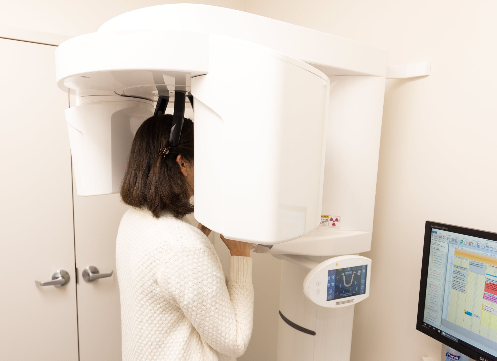
Experience the cutting-edge technology of dental cone beam computed tomography (CT) - a revolutionary tool that goes beyond regular x-rays. With just one scan, our state-of-the-art equipment produces stunning 3-D images of your teeth, soft tissues, nerve pathways, and bone. Get a comprehensive view of your dental health like never before, thanks to this groundbreaking technology.
Preparing for this procedure is simple. If there is a chance you might be pregnant, inform your doctor. Dress in loose and comfortable clothing and avoid wearing jewelry. It is possible that you will need to wear a gown.
What Is a CBCT Scan?
Advanced imaging technologies have significantly impacted the field of dentistry by improving the diagnosis and treatment of oral conditions. One notable technology is the Cone Beam Computed Tomography (CBCT) scan, which offers dentists and specialists detailed 3-dimensional images of a patient's oral structures in a non-invasive manner.
CBCT scan Applications
CBCT scans are widely used in dentistry for various purposes, including implant planning, oral surgery, and the evaluation of dental problems like impacted wisdom teeth, TMJ disorders, and root canal complications. The high-definition images produced by CBCT have made it an essential tool for providing customized and efficient dental care.
Before making a decision on whether CBCT Scans are suitable for you, there are some important facts you should be aware of.
Who Needs CBCT Scan?
Reimagine CBCT scans are utilized in modern dentistry to obtain precise images for diagnosing and planning dental treatments. Although they offer numerous advantages, CBCT scans are not always required for every dental patient. Dental professionals recommend CBCT scans based on individual indications.
- Dental Implant Planning: CBCT scans are necessary for patients who are considering dental implants because they provide a thorough view of the jawbone's structure, density, and proximity to vital structures. This information helps dentists determine the best placement for the implant, improving its long-term success and minimizing complications.
- Evaluation of Impacted Teeth: CBCT scans are used to assess impacted teeth, which are teeth that do not emerge correctly through the gum line. Whether it is impacted wisdom teeth or other molars, CBCT scans provide detailed visuals of the tooth's position and orientation, allowing dentists or oral surgeons to plan extractions safely and effectively.
- Diagnosis of TMJ Disorders: Temporomandibular joint (TMJ) disorders can result in discomfort and pain in the jaw joint. CBCT scans are used to assess the structure of the TMJ, detect any abnormalities, and assist in determining suitable treatment approaches.
- Evaluation of Root Canal Complications: CBCT scans are utilized in complex endodontic cases or when standard X-rays do not provide sufficient information. They aid endodontists in diagnosing problems such as root fractures, canal curvature, and persistent infections, resulting in more effective root canal treatments.
- Assessment of Pathologies and Tumors: CBCT scans are used to detect and assess oral pathologies, cysts, and tumors. They help dental professionals visualize these abnormalities' size, location, and extent, allowing for prompt planning of biopsies or referral to specialists for further evaluation.
- Orthodontic Treatment Planning: CBCT scans are used by orthodontists to help them create accurate treatment plans for patients who need orthodontic interventions. These scans provide a 3D visualization of the teeth and jaw relationships, allowing orthodontists to develop personalized treatment approaches, such as braces or Invisalign clear aligners, to achieve the best possible outcomes.
- Evaluation of Trauma: CBCT scans are utilized in cases of dental trauma or facial injuries to evaluate the extent of damage to the teeth, jawbone, and adjacent structures. This aids in formulating treatment strategies for restoring dental function and aesthetics.
CBCT scans offer valuable information but involve a slightly higher radiation dose than traditional dental X-rays. Dentists carefully evaluate the risk-benefit ratio before recommending CBCT scans and ensure that the benefits outweigh the potential risks, particularly for pregnant women and children. If you have additional inquiries about CBCT scans, please reach out to Artin Dental.
What Are The Advantages of CBCT Scan?
The incredible Cone Beam Computed Tomography (CBCT) scan revolutionizes dental diagnostics and treatment planning, unlocking a world of possibilities for dental professionals. Prepare to be amazed by the countless advantages of CBCT scans, as they become an essential tool for dental wizards everywhere. Brace yourself for the magic!
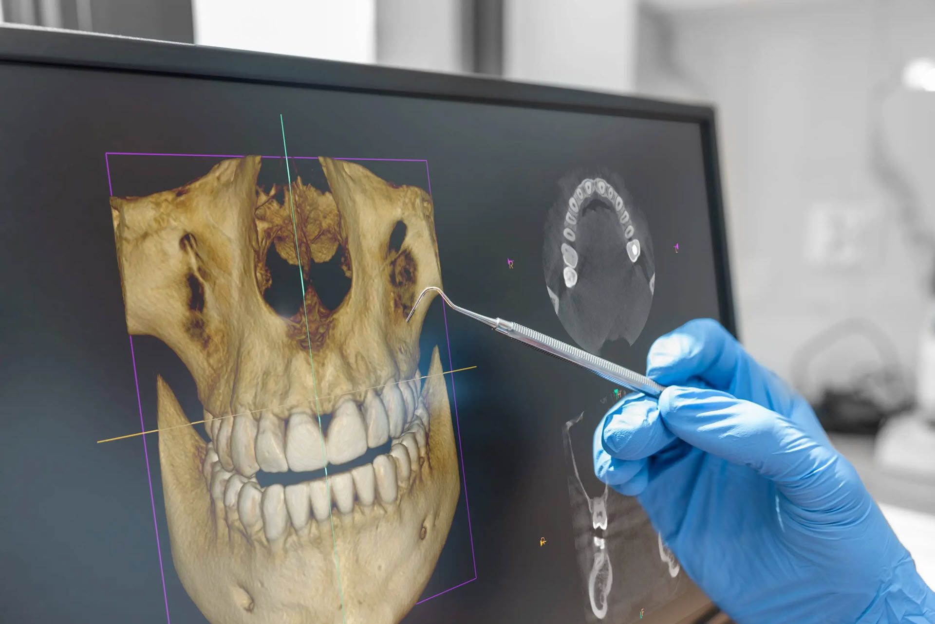
- Precise 3D Imaging: CBCT scans produce high-quality, three-dimensional images of the oral structures, providing a thorough view from various perspectives. This detailed visualization enables dental professionals to accurately evaluate dental anatomy, bone density, and the spatial relationship between teeth, nerves, and other important structures.
- Improved Diagnostic Capabilities: CBCT scans are an effective tool for diagnosing dental issues that may not be visible on traditional 2-dimensional X-rays. They help detect dental abnormalities, pathologies, and structural anomalies at an early stage, leading to more accurate and timely diagnoses.
- Treatment Planning: CBCT scans are utilized in dental practice to aid in treatment planning for a range of procedures, such as dental implants, oral surgeries, and orthodontic treatments. These scans provide dentists with detailed 3D images, allowing for a better understanding of the patient's anatomy and the development of customized treatment plans. This, in turn, enhances treatment accuracy and overall effectiveness.
- Reduced Need for Additional Imaging: CBCT scans are comprehensive and can provide a large amount of information, which often eliminates the need for multiple imaging studies. This means that dental professionals can minimize the use of additional X-rays and reduce patient exposure to radiation.
Addition Benefits of CBCT Scan
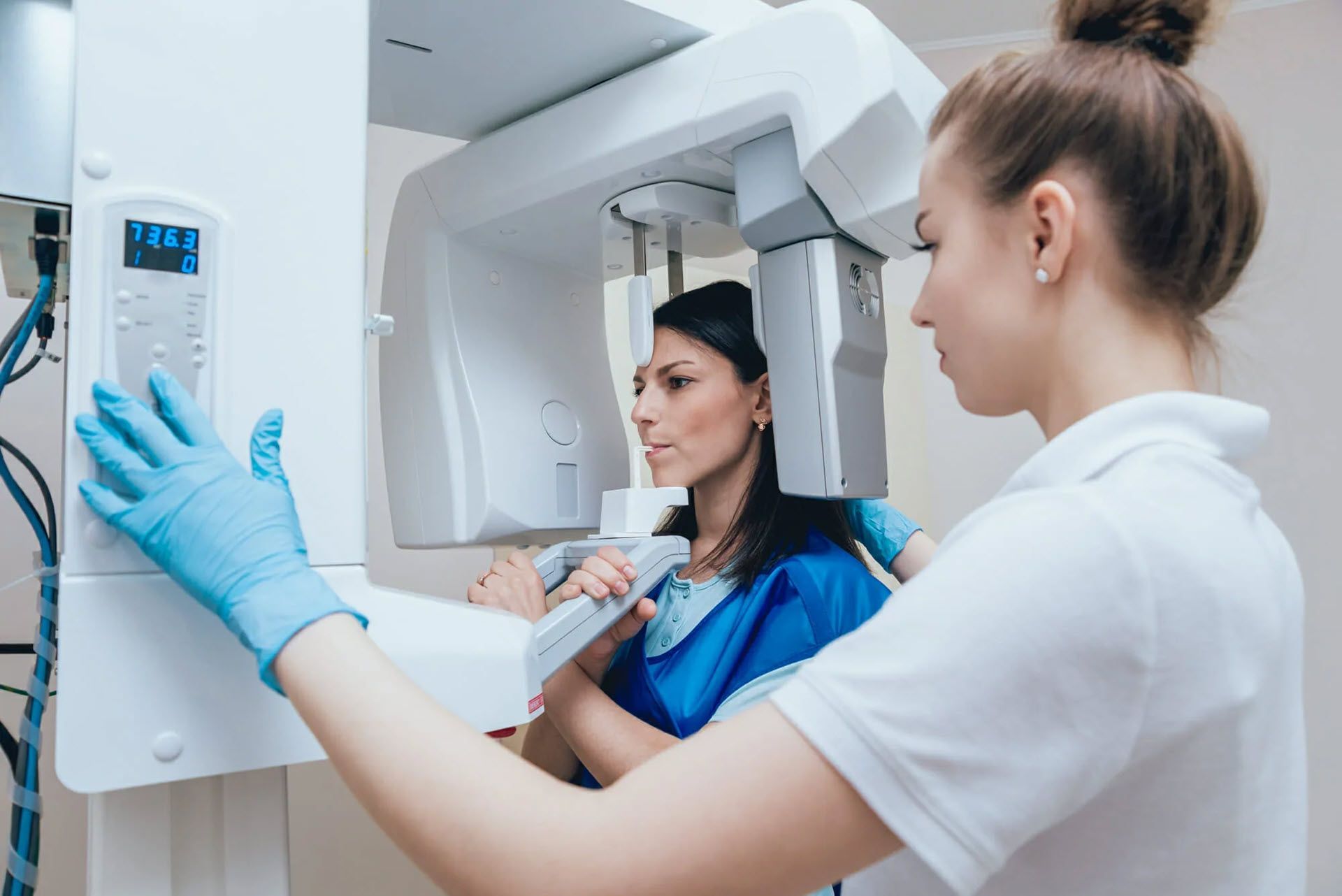
- Faster and Non-Invasive: CBCT scans are known for their efficiency, typically requiring only a few seconds to capture the necessary images. Unlike traditional medical CT scans, CBCT scans are non-invasive and do not require contrast dyes or extensive patient preparation.
- Greater Patient Comfort: Patients often report that CBCT scans are more comfortable than traditional CT scans. The scan is of short duration, and patients must stay still during the procedure, which helps reduce any discomfort or anxiety typically associated with it.
- Enhanced Implant Success: CBCT scans are used for dental implant planning as they provide crucial information about bone quality, quantity, and location, which helps ensure accurate implant placement. This plays a significant role in the success and durability of dental implants.
- Orthodontic Applications: CBCT scans are utilized in orthodontics to evaluate tooth and jaw relationships, detect impacted teeth, and facilitate effective treatment planning. They allow orthodontists to develop individualized treatment plans, resulting in enhanced patient outcomes.
- Visualization of Soft Tissues: CBCT scans provide visualization of dental structures as well as surrounding soft tissues, including muscles and nerves. This can be useful for evaluating conditions related TMJ disorders and other soft tissues.
- Enhanced Patient Education: CBCT scans enable effective communication between dental professionals and patients by providing clear, interactive visuals. These 3D images allow patients to better understand their dental conditions, explore potential treatment options, and actively engage in making decisions about their oral health.
CBCT scans have greatly impacted dental imaging and treatment, providing dentists and specialists with the ability to offer more precise and efficient care that is centered around the patient. The benefits of CBCT technology make it a valuable tool in modern dentistry, improving diagnostic accuracy, treatment results, and patient contentment. If you have any additional inquiries regarding CBCT Scans, please feel free to reach out to us.
Book Your Appointment
Book an appointment today with our doctors to discuss Dental Crowns
Artin Dental's CBCT Scan at Downtown Toronto
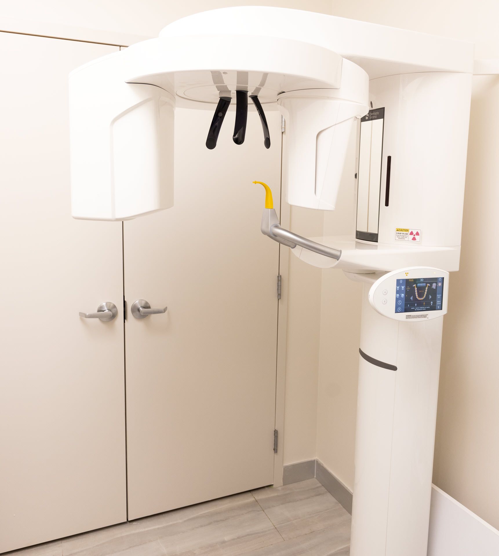
At Atrin Dental, we offer CBCT scans to help our patients receive the best dental care available. Our Cone Beam Computed Tomography (CBCT) scan is a 3D imaging system that allows us to see inside the teeth and surrounding structures in a precise way. By taking an accurate image of your mouth, we can diagnose and plan treatments more accurately.
If you’re interested in receiving a CBCT scan at our Downtown Toronto location, here are some steps to get you started:
1. Schedule an appointment - Contact us today to book an appointment for your CBCT scan. Please let us know if you have any medical conditions or medications that may affect the scan results.
2. Prepare for the appointment - Before your visit, drink plenty of water and avoid eating food for one hour prior to the appointment so that your stomach does not interfere with the scanning process.
3. Have your scan - During the procedure, you will be asked to hold a bite block between your teeth as part of the scanning process. The entire procedure takes no more than 10 minutes.
4. Get your results - Afterward, our expert team will review and interpret your images and provide you with detailed results about what was found during the scan along with treatment recommendations if needed. We will also discuss other imaging options if necessary, based on individual circumstances.
We look forward to helping you receive optimal dental care with our advanced CBCT Scan technology! Our dental clinic is conveniently located in the PATH of the Exchange Tower and First Canadian Place to serve you with CBCT dental scans. If you have any further inquiries or would like to schedule an appointment, please contact us today!
Frequently Asked Questions

Navigation
Services
Business Hours
- Mon, Wed, Thu
- -
- Tuesday
- -
- Friday
- -
- Sat - Sun
- Closed